
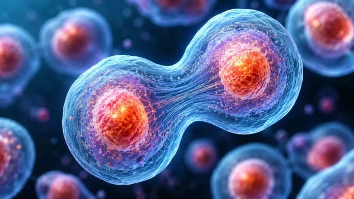

How Do Cells Divide?
Cells divide so living organisms can grow, replace damaged tissue, reproduce, and maintain normal body functions. Every new cell begins with information inherited from an existing cell, which means division must be carefully controlled so genetic material is copied and distributed correctly. In humans and many other organisms, the most familiar form of cell division is mitosis, which produces two genetically similar daughter cells. A second process called meiosis creates reproductive cells such as sperm and eggs and reduces the chromosome number by half. These two forms of division serve different purposes but both depend on accurate DNA replication and chromosome movement. Understanding how cells divide provides a foundation for learning about growth, healing, reproduction, genetics, aging, and diseases such as cancer.
Cell division does not happen randomly whenever a cell becomes large enough. Cells move through an organized sequence called the cell cycle, which includes periods of growth, DNA replication, preparation, and actual division. During this cycle, the cell checks whether conditions are suitable and whether its DNA has been copied correctly before moving forward. Specialized proteins act as control systems that can slow or stop the process when damage is detected. If these safeguards fail, cells may divide with damaged DNA or continue multiplying when they should stop. This is why cell-cycle regulation is such an important part of normal biology.
The process is especially impressive because DNA molecules are extremely long and must be duplicated before a cell divides. Human cells normally contain 46 chromosomes, and each chromosome must be copied so both new cells can receive a complete genetic set. During mitosis, duplicated chromosomes become tightly packed and are separated by microscopic structures that pull one copy toward each end of the cell. The cell then divides its cytoplasm and membrane, producing two separate daughter cells. Each daughter cell can continue living, performing specialized functions, or eventually dividing again depending on its role in the body. This constant cycle happens throughout many tissues every day.
This article explains how cell division works, including the cell cycle, DNA replication, mitosis, meiosis, cytokinesis, chromosomes, spindle fibers, and cell-cycle checkpoints. It also explores why cells divide, what happens when division goes wrong, how cancer develops, and why different cells divide at different rates. You will learn the major stages of mitosis and meiosis in a simple order that makes them easier to remember. The goal is to connect microscopic events inside cells with familiar processes such as growth, wound healing, and reproduction. Once the basic sequence is understood, cell division becomes much easier to visualize and remember.
Why Do Cells Need to Divide?
One major reason cells divide is growth. A human begins as a single fertilized cell, yet the adult body eventually contains trillions of cells with different structures and functions. That enormous increase is possible because cells repeatedly copy their DNA and divide into new cells. As development continues, many of those cells specialize into tissues such as muscle, skin, bone, blood, and nerves. Cell division therefore allows the body to become larger while preserving the genetic information needed by new cells. Without division, multicellular organisms could not grow beyond a single or very small number of cells.
Cell division is also essential for replacing old or damaged cells. Skin cells are continuously lost from the body, blood cells have limited lifespans, and cells lining the digestive tract experience constant wear. New cells must replace those that die so tissues can continue functioning properly. Bone marrow, for example, continually produces new blood cells through repeated cell division and differentiation. Wound healing also depends on nearby cells multiplying to replace tissue damaged by injury. This ability to renew tissues is one of the most important everyday functions of cell division.
Some organisms use cell division as a form of reproduction. Many bacteria reproduce by splitting into two cells through a process called binary fission, while certain single-celled eukaryotes also reproduce through forms of cell division. In these cases, one dividing cell can effectively create a new independent organism rather than simply contributing to a larger body. Multicellular organisms usually rely on more specialized reproductive processes, but cell division remains central to producing reproductive cells. Meiosis creates sperm and egg cells that can later combine during fertilization. Division is therefore closely connected with reproduction across many forms of life.
Cells also divide because becoming indefinitely larger would create biological problems. A very large cell would have difficulty moving materials efficiently between its center and outer membrane, and one set of genetic instructions might become insufficient for an enormous volume of cytoplasm. Dividing allows cells to maintain more manageable sizes and more efficient surface-area-to-volume relationships. Smaller cells can often exchange nutrients, gases, and waste products with their surroundings more effectively. Cell size is therefore not simply a matter of appearance but affects how efficiently a cell can operate. Division helps maintain dimensions that support normal cellular function.
What Is the Cell Cycle?
The cell cycle is the organized sequence through which a cell grows, copies its DNA, prepares for division, and eventually separates into daughter cells. It is commonly divided into interphase and the mitotic phase, although several smaller stages exist within those broader categories. Most of a cell’s life is actually spent in interphase rather than visibly dividing. During interphase, the cell grows, performs normal functions, and prepares its genetic material for duplication and separation. The cell cycle therefore includes much more than the few dramatic minutes when chromosomes can be seen moving. Understanding the full cycle explains how cells prepare for accurate division.
Interphase consists mainly of the G1, S, and G2 phases. During G1, the cell grows, produces proteins, and carries out many of its normal functions. During the S phase, DNA replication occurs and every chromosome is copied. During G2, the cell continues growing and prepares the structures and proteins needed for mitosis. These phases allow the cell to complete important tasks before it attempts to divide. Rushing directly into chromosome separation without preparation would dramatically increase the risk of genetic errors.
After interphase, the cell enters the mitotic phase, which includes mitosis and usually cytokinesis. Mitosis separates the duplicated chromosomes into two sets, while cytokinesis divides the cell’s cytoplasm and membrane. The exact timing and duration of each phase can vary widely depending on cell type and biological conditions. Some rapidly renewing cells move through the cycle regularly, while other cells may remain outside active division for long periods. Certain mature cells can even remain permanently in a nondividing state. The cell cycle is therefore flexible rather than identical in every tissue.
Cells do not move through the cycle without supervision. Checkpoints monitor DNA integrity, chromosome attachment, cell size, and whether conditions are suitable for division. If a problem is found, the cycle can pause while the cell attempts repair. When damage is too severe, the cell may activate programmed cell death instead of passing damaged genetic material to new cells. These checkpoints help protect organisms from dangerous mutations and abnormal growth. Failures in these control systems are strongly connected with diseases such as cancer.
What Happens During Interphase?
Interphase is sometimes mistakenly described as a resting period, but cells are highly active throughout it. During G1, the cell increases in size, creates new proteins, produces organelles, and performs its specialized functions. A liver cell, for example, continues participating in metabolism while also preparing for possible division. Nutrients and energy are used to build the materials needed for future growth. The cell also evaluates whether conditions are favorable enough to continue through the cycle. This stage can vary greatly in length depending on the type of cell.
The S phase is especially important because this is when DNA is replicated. Before replication, each chromosome contains one DNA molecule, but after the S phase each chromosome has been duplicated. The two identical copies remain connected and are known as sister chromatids. These chromatids contain almost identical genetic information and must later be separated so each daughter cell receives one copy. Accurate DNA replication is therefore essential for genetic continuity. Errors occurring during this phase can become mutations if they are not repaired.
During G2, the cell checks the duplicated DNA and prepares for mitosis. Proteins involved in chromosome movement and spindle formation are produced, and organelles continue being duplicated or reorganized. The cell also accumulates enough energy and resources to complete division successfully. A checkpoint near the end of G2 helps determine whether DNA replication was completed correctly. If serious problems remain, progression into mitosis can be delayed. This stage ensures that the cell does not begin chromosome separation before it is ready.
Some cells leave the active cycle and enter a state known as G0. In this state, cells perform their normal specialized functions without preparing immediately for division. Certain cells can return from G0 and start dividing again when the body needs them, while others remain in this state for extremely long periods. Many mature nerve cells, for example, rarely divide under normal conditions. This helps explain why different tissues have very different abilities to regenerate after injury. The decision to divide therefore depends strongly on cell type and biological signals.
How Is DNA Copied Before Cell Division?
DNA must be copied before a cell divides so each daughter cell can receive a complete set of genetic instructions. The process begins when the DNA double helix unwinds and the two strands separate in specific regions. Each original strand then acts as a template for building a complementary new strand. Special enzymes recognize the sequence of bases and add matching nucleotides according to base-pairing rules. Adenine pairs with thymine, while cytosine pairs with guanine. This allows the genetic sequence to be reproduced with extremely high accuracy.
An enzyme called DNA polymerase plays a central role in creating the new strands. It moves along the template DNA and adds complementary nucleotides one after another. Other enzymes help unwind DNA, stabilize separated strands, and connect newly synthesized sections. Because chromosomes contain millions of DNA bases, replication begins at many locations simultaneously rather than starting at only one end. This greatly increases the speed of the process. The entire genome can therefore be copied within a reasonable amount of time.
DNA replication also includes proofreading and repair mechanisms. DNA polymerases can detect many incorrectly paired bases and correct them before replication continues. Additional repair proteins inspect newly copied DNA and remove mistakes that escaped the first proofreading step. These systems are remarkably effective, although they cannot eliminate every error. A small number of mistakes may remain and become permanent mutations. This is one reason spontaneous genetic changes can appear during normal cell division.
After replication, each chromosome consists of two sister chromatids joined together at a region called the centromere. These chromatids remain connected until the appropriate stage of mitosis. Their temporary attachment ensures that the cell can move both copies together before separating them accurately. Proteins surrounding the centromere later form structures that interact with spindle fibers. This connection is essential for pulling chromosomes toward opposite ends of the dividing cell. DNA replication and chromosome separation are therefore tightly coordinated parts of the same process.
What Is Mitosis?
Mitosis is the process that separates duplicated chromosomes into two genetically similar sets. It is used for growth, tissue repair, replacement of old cells, and certain forms of asexual reproduction. Mitosis does not directly divide the cytoplasm; instead, it organizes and separates the nuclear genetic material before cytokinesis finishes the division. In humans, a typical body cell entering mitosis has already duplicated its 46 chromosomes. Each daughter cell normally ends up with the same chromosome number as the original cell. This genetic consistency is one of the main purposes of mitotic division.
Mitosis is commonly divided into several stages known as prophase, metaphase, anaphase, and telophase. Some textbooks also describe prometaphase as a separate stage between prophase and metaphase. These stages are not completely isolated events but parts of one continuous sequence. Chromosomes gradually condense, align, separate, and become enclosed within new nuclei. Scientists divide the process into named stages because doing so makes the events easier to study and understand. In a real cell, one stage smoothly transitions into the next.
During mitosis, a structure called the mitotic spindle plays a central role in chromosome movement. The spindle is made mainly from microtubules that extend from opposite sides of the cell. These fibers attach to chromosome structures called kinetochores and help position chromosomes before separation. Once every chromosome is properly connected, spindle fibers shorten or move in coordinated ways to separate sister chromatids. Accurate spindle attachment is essential because improper separation can produce cells with abnormal chromosome numbers. Checkpoints therefore monitor whether chromosomes are correctly connected before the cell proceeds.
Mitosis is extremely reliable, but mistakes can occur. A chromosome may fail to separate correctly, or DNA damage may pass through checkpoints without being repaired. Such errors can produce genetically abnormal daughter cells and may contribute to disease if they affect important genes. The body has multiple systems for detecting abnormal cells and preventing them from continuing to divide. However, some cells eventually escape these controls. Cancer is one major example of what can happen when normal cell-division regulation breaks down.
What Happens During Prophase?
Prophase is the first major visible stage of mitosis. During this stage, the long, loose DNA molecules present during interphase begin condensing into compact chromosomes. Condensation is important because tightly packed chromosomes are easier for the cell to move without tangling or breaking. Each visible chromosome consists of two sister chromatids that were created during DNA replication. The chromatids remain connected at their centromere while the cell prepares for separation. This is why chromosomes appear thicker and more distinct under a microscope during mitosis.
The nucleolus, a structure inside the nucleus involved in ribosome production, begins disappearing during prophase. At the same time, centrosomes that were previously duplicated move toward opposite sides of the cell. These structures help organize the microtubules that will form the mitotic spindle. As they move apart, spindle fibers begin extending through the cell. Their arrangement creates the framework needed for chromosome positioning. Without this coordinated organization, chromosome separation would be highly unreliable.
As prophase progresses into prometaphase, the nuclear envelope breaks down. This allows spindle microtubules to reach the chromosomes directly. Specialized protein structures called kinetochores are located around the centromere region of each chromosome. Microtubules attach to these kinetochores and begin moving chromosomes toward the center of the cell. Different spindle fibers may push, pull, or stabilize chromosomes during this process. The goal is to establish proper connections from opposite sides of the cell.
Prophase therefore prepares nearly every structure needed for successful chromosome separation. DNA becomes compact, the nuclear boundary is removed, spindle fibers form, and chromosomes establish connections with the spindle. These events transform a cell from its normal interphase arrangement into a highly organized dividing system. Although chromosome movement is not yet complete, the machinery responsible for later separation is already in place. Errors during this stage can disrupt later steps and activate cell-cycle checkpoints. Accurate preparation during prophase is therefore essential for successful mitosis.
What Happens During Metaphase?
During metaphase, duplicated chromosomes become aligned across the middle of the cell. This central region is often called the metaphase plate, although it is not a physical plate or membrane. Each chromosome is positioned so one sister chromatid is connected to spindle fibers originating from one side of the cell and the other chromatid is connected to the opposite side. This balanced arrangement prepares the two copies for separation. Proper alignment is visually striking when viewed under a microscope. It represents one of the most carefully controlled stages of mitosis.
The spindle checkpoint is especially active during metaphase. The cell must confirm that every chromosome is correctly attached before separation begins. If one chromosome lacks proper attachment, the cell can delay progression and continue attempting to fix the problem. This prevents one daughter cell from receiving an extra chromosome while the other receives too few. Accurate chromosome number is essential because large imbalances can alter many genes at once. The checkpoint therefore acts as an important safeguard against genetic instability.
Tension across sister chromatids helps the cell determine whether attachments are correct. When opposite spindle fibers pull properly, the chromosome experiences balanced force from both sides. Incorrect attachment produces abnormal tension and can trigger corrective mechanisms. The cell repeatedly adjusts these connections until the chromosomes are arranged correctly. This microscopic quality-control system demonstrates how carefully chromosome segregation is regulated. Successful mitosis depends on much more than simply pulling chromosomes apart.
Once every chromosome satisfies the spindle checkpoint, the cell receives biochemical signals that allow anaphase to begin. Proteins holding sister chromatids together are broken down, releasing the duplicated chromosome copies. At this moment, each chromatid becomes considered an individual chromosome. The cell is now ready to move one complete chromosome set toward each end. Metaphase therefore represents the final verification stage before irreversible chromosome separation. The next phase transforms one aligned genetic set into two distinct groups.
What Happens During Anaphase?
Anaphase begins when sister chromatids separate from one another. Proteins that previously held the chromatids together are cleaved, allowing each copy to move independently. Spindle fibers attached to kinetochores guide the chromosomes toward opposite poles of the cell. One complete copy of every chromosome moves toward one side, while the matching copies move toward the other. This separation ensures that the future daughter cells receive nearly identical genetic information. The movement is rapid compared with much of the rest of the cell cycle.
Different microtubules contribute to chromosome movement in coordinated ways. Some shorten as chromosomes move toward the poles, while other spindle fibers help push the two ends of the cell farther apart. Motor proteins also contribute mechanical force along microtubules. The result is an organized separation rather than chromosomes simply floating apart randomly. Cellular machinery precisely controls both direction and timing. This complexity allows dozens of chromosomes to be distributed accurately at the same time.
As anaphase progresses, the cell becomes visibly elongated. The increasing distance between chromosome groups helps create enough space for two new nuclei to form later. Each group contains one copy of every chromosome that was duplicated before mitosis. In a normal human body cell, each end receives a full set of 46 chromosomes. This maintains the chromosome number from one generation of cells to the next. Mitosis therefore preserves genetic continuity throughout growth and tissue renewal.
Errors during anaphase can produce serious consequences if chromosomes move incorrectly. A phenomenon called nondisjunction occurs when chromosomes or chromatids fail to separate properly. The resulting daughter cells may contain abnormal numbers of chromosomes, a condition known as aneuploidy. Cells often detect severe abnormalities and stop dividing, but some abnormal cells can survive. Chromosome instability is frequently observed in cancer cells. Accurate anaphase therefore remains one of the most critical steps in successful cell division.
What Happens During Telophase?
Telophase begins after chromosomes have reached opposite ends of the dividing cell. Once separated, the chromosomes start losing their tightly condensed appearance and gradually return to a more relaxed form called chromatin. This makes the DNA easier for the new cells to access once normal gene activity resumes. The dramatic chromosome structures visible during earlier mitosis therefore begin disappearing. Cell division is now approaching completion. The genetic material has already been successfully separated into two groups.
New nuclear envelopes begin forming around each chromosome set. Proteins and membrane components reconstruct the nuclear boundary so each daughter cell can have its own nucleus. Nucleoli also begin reappearing as normal nuclear functions restart. The spindle fibers that moved chromosomes are dismantled because they are no longer needed. These changes reverse many events that occurred during prophase. By the end of telophase, two distinct nuclei exist within what may still temporarily be one continuous cell.
Chromosomes continue becoming less condensed throughout telophase. This transition is important because tightly packed mitotic chromosomes are not ideal for normal transcription and gene expression. Cells need DNA in a more accessible state to resume protein production and everyday biological functions. The daughter nuclei therefore transition from division mode back toward normal cellular activity. The cell also begins preparing its cytoplasm for complete physical separation. In many animal cells, cytokinesis is already underway before telophase has completely finished.
Telophase does not create genetically new information; instead, it organizes the duplicated DNA into two stable nuclear compartments. Each nucleus normally contains the same chromosome number and nearly identical genetic sequence as the original parent nucleus. Small differences may exist if mutations occurred during replication or division, but the intended outcome is genetic consistency. Once nuclear organization is complete, cytokinesis can fully separate the two cells. Mitosis and cytokinesis therefore work together as consecutive parts of cell division. Their completion marks the beginning of two new daughter cells.
What Is Cytokinesis?
Cytokinesis is the physical division of the cell’s cytoplasm into two separate daughter cells. Mitosis organizes and separates the chromosomes, while cytokinesis completes the division of the rest of the cell. In animal cells, cytokinesis usually begins when the cell membrane starts pinching inward around the middle. A ring of proteins beneath the membrane contracts and gradually narrows the connection between the two sides. This indentation is called a cleavage furrow. Eventually, the membrane separates completely and produces two independent cells.
The contractile ring involved in animal-cell cytokinesis contains proteins such as actin and myosin. These proteins are also involved in other forms of movement and contraction within cells and tissues. As the ring tightens, it pulls the membrane inward while organelles and cytoplasmic materials are distributed between the developing daughter cells. The process must be coordinated with chromosome separation so the membrane does not divide through the genetic material. Cellular signaling helps ensure that the cleavage furrow forms in the correct location. Proper timing is therefore essential for safe division.
Plant cells cannot divide by simply pinching inward because they are surrounded by rigid cell walls. Instead, vesicles gather near the center of the dividing plant cell and fuse to form a structure called the cell plate. The cell plate expands outward until it connects with the existing cell membrane and wall. New cell-wall material is deposited between the two daughter cells. This creates a permanent boundary separating them. Plant cytokinesis therefore achieves the same goal through a different mechanical process.
After cytokinesis, each daughter cell enters a new stage of life. It may grow, perform specialized functions, enter G0, or begin another cell cycle depending on signals and tissue requirements. The two cells are genetically very similar because they received chromosome copies from the same parent cell. Their future behavior, however, can become different if they activate different genes or receive different environmental signals. Cell division therefore produces genetic continuity without requiring every daughter cell to remain functionally identical. Development and tissue organization depend heavily on this ability.
What Is Meiosis?
Meiosis is a specialized form of cell division used to produce reproductive cells. Unlike mitosis, which normally produces two genetically similar cells, meiosis produces cells containing half the original chromosome number. In humans, body cells typically contain 46 chromosomes, while sperm and egg cells contain 23. This reduction is necessary because fertilization later combines one sperm and one egg. Their chromosome sets join to restore the normal total of 46. Without meiosis, chromosome numbers would double with every generation.
Meiosis involves two consecutive rounds of division known as meiosis I and meiosis II. DNA is copied only once before these two divisions begin. During meiosis I, homologous chromosome pairs are separated from one another. During meiosis II, sister chromatids separate in a process that resembles mitosis. The overall result is usually four haploid cells rather than two diploid cells. Each final cell contains one chromosome from every original homologous pair.
One of the most important features of meiosis is that it generates genetic variation. Homologous chromosomes inherited from the two biological parents can exchange corresponding DNA segments through a process called crossing over. Chromosome pairs also align independently during meiosis I, creating different possible combinations in reproductive cells. These processes ensure that sperm and egg cells are genetically different from one another. Fertilization adds another layer of variation because any one sperm may combine with any one egg. Sexual reproduction therefore creates enormous genetic diversity.
Meiosis also involves strict chromosome control because errors can affect future offspring. If chromosomes fail to separate correctly, reproductive cells may receive too many or too few chromosomes. After fertilization, this can produce embryos with chromosome abnormalities. Some abnormalities prevent normal development, while others can result in recognizable genetic conditions. The risk of certain separation errors can change with parental age and other biological factors. Accurate meiosis is therefore essential for maintaining chromosome number across generations.
How Is Meiosis Different From Mitosis?
The main difference is purpose. Mitosis produces new body cells for growth, repair, and replacement, while meiosis produces reproductive cells needed for sexual reproduction. A skin cell dividing to replace damaged tissue uses mitosis, whereas cells developing into sperm or eggs undergo meiosis. These different goals require different chromosome outcomes. Mitosis preserves chromosome number, while meiosis reduces it by half. This distinction prevents chromosome doubling during fertilization.
Mitosis involves one main nuclear division after DNA replication. Meiosis involves two consecutive divisions after one round of DNA replication. As a result, mitosis usually creates two daughter cells, while meiosis typically produces four cells. The daughter cells from mitosis are usually genetically similar to the parent cell. Cells produced through meiosis are genetically different because of crossing over and independent chromosome assortment. This genetic variation is one of the defining features of sexual reproduction.
Chromosome pairing also differs between the two processes. During meiosis I, homologous chromosomes pair together and may exchange DNA. Homologous pairs consist of corresponding chromosomes inherited from each biological parent. Mitosis does not normally involve this pairing or crossing-over process. Instead, duplicated chromosomes line up individually and sister chromatids are separated directly. This difference explains why meiosis can rearrange inherited genetic combinations while mitosis generally preserves them.
The final chromosome number also makes the distinction easy to remember. Human mitosis normally begins with a diploid cell containing 46 chromosomes and produces daughter cells that also contain 46. Human meiosis begins with a diploid precursor cell but ultimately produces haploid cells containing 23 chromosomes. Fertilization combines two haploid cells and restores diploid chromosome number in the new embryo. Mitosis then takes over to produce the enormous number of cells needed for development. Both processes are therefore essential but serve very different biological roles.
What Are Chromosomes Doing During Cell Division?
Chromosomes are organized packages of DNA and associated proteins. Before division, DNA exists in a relatively loose form that allows genes to be accessed and used by the cell. When mitosis begins, the DNA becomes highly condensed so chromosomes can be moved safely. This packaging prevents long DNA molecules from becoming tangled during separation. Each duplicated chromosome contains two sister chromatids. These copies remain connected until the correct stage of division.
Centromeres play a central role in chromosome movement. They are specialized chromosome regions where protein structures called kinetochores form. Spindle microtubules attach to kinetochores and generate the forces needed to position and separate chromosomes. The centromere therefore acts as an important connection point between genetic material and the machinery of division. Problems affecting centromeres or kinetochore attachment can lead to chromosome segregation errors. Accurate attachment is closely monitored by the cell.
Chromosome condensation is temporary. Once separation is complete and daughter nuclei form, chromosomes relax into chromatin again. This change allows transcription machinery to access genes and resume ordinary cellular functions. Cells therefore switch DNA between more compact and more accessible configurations depending on what they are doing. Division favors safe transport, while normal cell activity favors accessibility. Chromosome structure is therefore dynamic rather than fixed.
Chromosomes also carry information beyond individual genes. They contain regulatory regions, centromeres, telomeres, and other sequences important for stability and organization. Telomeres at chromosome ends help protect DNA from being mistaken for broken genetic material. Each round of DNA replication creates challenges for maintaining telomere length, which is connected with cellular aging and division limits in many cell types. Cancer cells can sometimes activate mechanisms that maintain telomeres more effectively. Chromosome biology is therefore deeply connected with both normal division and disease.
What Controls the Cell Cycle?
The cell cycle is controlled by networks of regulatory proteins that determine whether a cell should move from one stage to the next. Important proteins include cyclins and cyclin-dependent kinases, often abbreviated as CDKs. Cyclin levels rise and fall during different phases, activating CDKs at appropriate times. These signals help coordinate DNA replication, chromosome separation, and other major events. The process resembles a series of molecular switches rather than one continuous automatic timer. Cells therefore divide only when the necessary regulatory conditions are met.
Several checkpoints monitor progress through the cycle. One important checkpoint occurs before DNA replication and asks whether the cell is large enough and conditions are favorable. Another checkpoint before mitosis checks whether DNA has been replicated completely and whether serious damage remains. The spindle checkpoint during mitosis confirms that chromosomes are properly attached before separation begins. Each checkpoint protects against a different type of error. Together they greatly improve the reliability of cell division.
Proteins involved in tumor suppression also help prevent damaged cells from dividing. One famous example is p53, which can respond to DNA damage by slowing the cell cycle and activating repair pathways. If the damage cannot be repaired, p53 may contribute to programmed cell death. This prevents severely damaged genetic material from being passed to daughter cells. Mutations affecting such control proteins can allow abnormal cells to survive and continue dividing. These failures are common features of many cancers.
External signals also influence whether cells divide. Growth factors, hormones, nutrients, contact with neighboring cells, and tissue conditions can all affect cell-cycle activity. Normal cells generally respond to these signals and stop dividing when growth is unnecessary. Cancer cells often become less responsive to normal growth controls and may produce their own signals or ignore inhibitory messages. This difference helps explain why tumors can continue expanding even when surrounding tissues do not need more cells. Cell division is therefore controlled by both internal checkpoints and external communication.
Why Do Some Cells Divide Faster Than Others?
Different tissues have different replacement needs, so their cells divide at different rates. Skin and intestinal lining cells experience frequent wear and must be replaced regularly. Bone marrow also produces enormous numbers of new blood cells because many circulating blood cells have limited lifespans. These tissues therefore contain populations of rapidly dividing cells or stem cells that continually generate replacements. Their high division rates are essential for maintaining normal body function. If production slows dramatically, tissue performance can deteriorate quickly.
Other cells divide much less frequently. Mature nerve cells in the brain and spinal cord generally have very limited ability to re-enter the cell cycle under normal conditions. Certain muscle cells also divide poorly once fully specialized. This is one reason injuries to these tissues can be difficult to repair completely. Instead of replacing lost cells easily, the body may rely on scar formation or limited regenerative mechanisms. Tissue regeneration therefore depends strongly on the division capacity of its cells.
Stem cells provide an important solution in many tissues. These cells can divide while maintaining the ability to produce more specialized daughter cells. One daughter cell may remain a stem cell, while another begins differentiating into a mature cell type. This balance allows tissues to maintain a long-term source of replacement cells. Bone marrow, skin, and intestinal tissue all depend on specialized stem-cell populations. Stem cells therefore connect cell division directly with tissue renewal.
Division rates can also change after injury or in response to hormones and growth signals. Cells that normally divide slowly may become more active when tissue repair is needed. Once healing is complete, inhibitory signals help reduce division again. Problems arise when cells ignore these signals and continue multiplying excessively. Cancer represents an extreme example of uncontrolled division. Normal tissue health therefore depends on both the ability to divide and the ability to stop dividing.
What Happens When Cell Division Goes Wrong?
Errors can occur during DNA replication, chromosome separation, or cell-cycle regulation. If a chromosome is copied incorrectly, mutations may appear in the daughter cells. If chromosomes fail to separate properly, one cell may receive too many copies while another receives too few. Some of these abnormalities cause immediate cell death, while others may remain unnoticed. The biological effect depends on which genes or chromosome regions are affected. Cell-division errors are therefore a major source of genetic variation and disease.
Many abnormal cells are eliminated by protective mechanisms. Checkpoints can stop division when DNA damage is detected, and programmed cell death can remove cells that cannot be repaired safely. Immune cells may also recognize and destroy certain abnormal cells. These systems work continuously throughout life. Most potentially dangerous cellular errors therefore never develop into disease. Problems arise when abnormal cells accumulate additional changes that allow them to escape control.
Chromosome-separation errors during meiosis can affect reproduction. If sperm or egg cells contain abnormal chromosome numbers, fertilization may produce an embryo with too many or too few chromosomes. Some chromosome abnormalities are incompatible with continued development, while others can lead to genetic conditions. These errors demonstrate why accurate spindle function and chromosome pairing are essential during meiosis. Reproductive cell division therefore requires particularly precise chromosome management. Even one separation error can affect every cell in the resulting organism.
Errors in mitotic control can contribute to tumors. A cell that acquires mutations allowing it to bypass checkpoints may continue dividing despite DNA damage. Additional mutations can give the cell further advantages, such as resistance to programmed death or the ability to stimulate blood-vessel growth. Over time, descendants of that abnormal cell can form a tumor. Cancer therefore develops through a combination of genetic changes and failures in cell-cycle regulation. Abnormal division is not the only feature of cancer, but it is one of its central characteristics.
How Does Cancer Relate to Cell Division?
Cancer develops when cells acquire changes that disrupt the normal controls over growth and division. Healthy cells usually divide only when signals indicate that new cells are needed. They also respond to checkpoints that prevent damaged DNA from being copied. Cancer cells can gradually lose these controls because mutations affect genes responsible for regulating the cell cycle. As these changes accumulate, cells may continue multiplying even when the body does not need more of them. This uncontrolled growth can eventually produce a tumor.
Some cancer-related genes normally encourage cell division when growth is appropriate. When these genes become abnormally activated, they can behave like stuck accelerators and push cells toward continuous proliferation. Other genes normally suppress tumors by slowing division, repairing DNA, or triggering cell death. Losing the function of these genes removes important biological brakes. Cancer often develops after several different control pathways have been disrupted. One mutation alone is usually not sufficient to create a fully malignant cell.
Cancer cells can also become genetically unstable. Because their checkpoints and DNA-repair systems may be impaired, additional mutations and chromosome abnormalities accumulate more quickly. This creates a population of tumor cells that may differ genetically from one another. Some cells can become more resistant to treatment, better at invading tissue, or more capable of spreading to distant organs. Tumor evolution therefore resembles a rapid form of cellular selection. Abnormal cell division continually generates new variation within the cancer.
Many cancer treatments target dividing cells. Chemotherapy drugs often interfere with DNA replication, chromosome separation, or other processes needed for rapid division. This can damage cancer cells but may also affect healthy tissues that normally divide quickly, such as hair follicles and the digestive lining. These effects help explain some common treatment side effects. Modern therapies increasingly try to target specific molecular abnormalities in cancer cells while protecting normal tissue. Understanding cell division therefore has direct importance for cancer diagnosis and treatment.
Why Is Cell Division Important for Wound Healing?
When tissue is injured, damaged or dead cells must be replaced. Nearby cells and specialized stem cells receive chemical signals that stimulate them to enter the cell cycle and begin dividing. New cells gradually fill the damaged region and help restore tissue structure. Blood vessels may also grow into the area to supply oxygen and nutrients. This coordinated response involves cell division, migration, inflammation, and extracellular-matrix production. Healing therefore depends on much more than simply closing the visible surface of a wound.
Skin provides a clear example. Cells near a cut begin multiplying and moving across the damaged area. Fibroblasts produce structural proteins that help rebuild connective tissue, while epithelial cells restore the protective surface. Immune cells remove bacteria and damaged material during earlier stages of healing. As repair progresses, cell division gradually slows. The tissue then enters remodeling stages that can continue long after the wound appears closed.
Different tissues have different healing capacities because their cells divide at different rates. Skin and liver tissue can regenerate relatively effectively, while certain nerve and heart cells have much more limited regenerative potential. Serious damage to poorly dividing tissues may therefore produce permanent loss of function or scar formation. Researchers are studying stem cells and regenerative medicine in hopes of improving repair in these tissues. Cell-division biology is central to these efforts. Understanding how to stimulate safe regeneration without causing uncontrolled growth is a major medical challenge.
Healing also requires division to stop at the correct time. If cells continued multiplying indefinitely after a wound was repaired, excess tissue could develop. Chemical signals and contact with neighboring cells normally help switch off the proliferative response. Problems with these signals can contribute to abnormal scarring or uncontrolled growth. Effective healing therefore depends on a carefully timed increase and decrease in cell division. The ability to stop is just as important as the ability to start.
Can Cells Divide Forever?
Most normal human body cells cannot divide indefinitely. After a certain number of divisions, many cells enter a state in which they no longer actively divide. One reason is that chromosome ends contain protective structures called telomeres, which gradually shorten during repeated DNA replication in many cell types. When telomeres become critically short, cells can activate pathways that stop further division. This acts as one protective mechanism against genetic instability. The phenomenon contributes to what researchers call cellular senescence.
Stem cells and certain other cell populations possess mechanisms that maintain telomeres more effectively. An enzyme called telomerase can add DNA sequences back to chromosome ends and slow telomere shortening. This allows selected cells to divide many more times than typical mature body cells. Reproductive cells and some stem cells use telomerase because long-term division is necessary for their biological roles. However, unlimited division must still be carefully regulated. Excessive proliferative capacity can become dangerous.
Many cancer cells activate telomerase or alternative mechanisms that maintain chromosome ends. This helps them avoid one of the natural barriers that normally limits repeated cell division. As a result, cancer cells may continue dividing far beyond the lifespan of ordinary cells. Telomere maintenance is therefore one feature that contributes to cellular immortality in many tumors. Researchers have investigated whether disrupting these mechanisms could provide useful cancer treatments. The challenge is targeting cancer cells without damaging normal stem-cell populations that also rely on telomere maintenance.
Cell division limits are connected with aging but do not explain aging by themselves. Organisms age because of many interacting processes involving DNA damage, protein changes, metabolism, immune function, inflammation, epigenetics, and tissue regeneration. Cellular senescence is one part of this broader picture. Cells that stop dividing can also release signals that influence neighboring tissues. Researchers continue studying whether removing or modifying senescent cells could improve certain age-related conditions. Cell-cycle biology therefore remains important far beyond basic growth and reproduction.
Final Thoughts on How Cells Divide
Understanding how cells divide begins with the cell cycle, an organized sequence that allows cells to grow, copy their DNA, check for errors, and eventually separate. Interphase prepares the cell by increasing its size and duplicating every chromosome. Mitosis then organizes and separates those duplicated chromosomes into two groups. Cytokinesis divides the remaining cytoplasm and cell membrane. The result is usually two daughter cells carrying nearly identical genetic information. This process supports growth, tissue repair, and replacement throughout the body.
Mitosis itself follows an orderly progression through prophase, metaphase, anaphase, and telophase. Chromosomes condense, align at the center of the cell, separate toward opposite poles, and become enclosed within new nuclei. Spindle fibers and kinetochores provide the mechanical system that moves chromosomes accurately. Checkpoints monitor the process and prevent separation when major problems are detected. These safeguards are essential because chromosome mistakes can affect many genes at once. Accurate mitosis therefore preserves genetic stability from one cell generation to the next.
Meiosis serves a different purpose by producing reproductive cells with half the normal chromosome number. It involves two divisions and creates genetic variation through chromosome assortment and crossing over. Sperm and egg cells produced through meiosis contain 23 chromosomes in humans, allowing fertilization to restore the normal total of 46. This process prevents chromosome numbers from doubling across generations. It also ensures that offspring receive unique genetic combinations. Meiosis therefore connects cell division directly with inheritance and evolution.
Cell division must always remain balanced with cell-cycle control. Too little division can impair growth and tissue repair, while too much uncontrolled division can contribute to cancer. DNA-repair systems, checkpoints, external growth signals, and programmed cell death work together to keep the process safe. Scientists continue studying cell division because it influences development, aging, fertility, regeneration, and many diseases. The process may occur on a microscopic scale, but its effects determine how entire organisms grow and survive. Cell division is therefore one of the most fundamental processes in biology.
Frequently Asked Questions
What are the main stages of cell division?
The major mitotic stages are prophase, metaphase, anaphase, and telophase, followed by cytokinesis. Before mitosis begins, the cell passes through interphase, where it grows and copies its DNA.
Why do cells divide?
Cells divide to support growth, replace old or damaged cells, heal tissues, and allow reproduction. Division also helps cells maintain manageable sizes and efficient internal transport.
What is the difference between mitosis and meiosis?
Mitosis produces two genetically similar body cells and preserves chromosome number. Meiosis produces reproductive cells, reduces chromosome number by half, and creates genetic variation.
What happens to DNA before a cell divides?
DNA is replicated during the S phase of interphase. Each chromosome becomes two sister chromatids so the dividing cell can provide one complete chromosome copy to each daughter cell.
What happens if cell division goes wrong?
Errors can produce mutations, abnormal chromosome numbers, damaged cells, infertility, developmental conditions, or cancer. Cell-cycle checkpoints and repair systems normally help prevent many of these problems.